
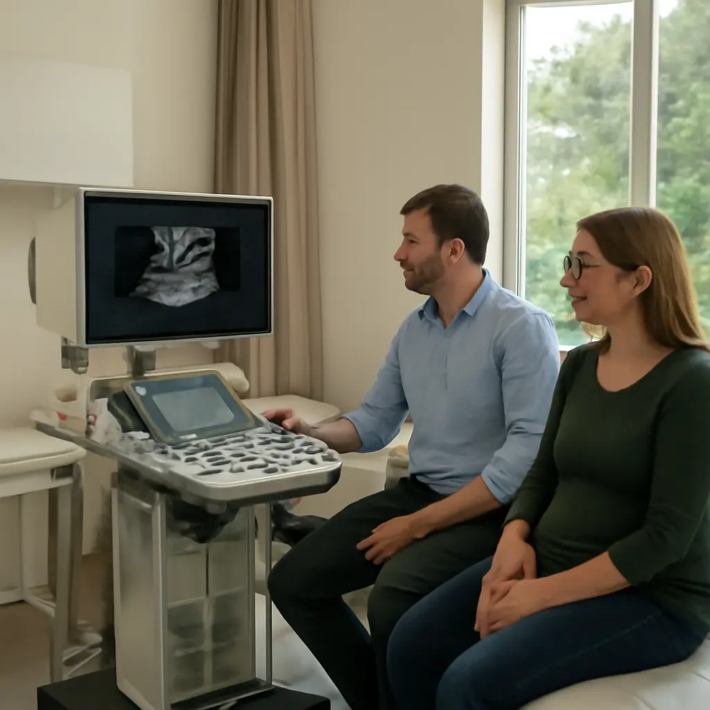
Prenatal screening: Inside the rooms where early detection guides care
Prenatal screening is the process of estimating the chance that a pregnancy has certain chromosome conditions or structural differences; it does not, by itself, diagnose them. In screening rooms, ultrasound imaging, blood tests, careful counseling, and—when needed—diagnostic testing help families and clinicians understand what to do next. The stakes are significant: the World Health Organization estimates that congenital disorders affect about 6% of babies worldwide, while the U.S. Centers for Disease Control and Prevention reports that birth defects affect roughly one in every 33 babies born in the United States. Screening can help identify some concerns earlier, support timely evaluation and treatment planning, and prepare families, while also requiring clear communication about uncertainty, choices, and limits.
Prenatal screening and early detection
Prenatal screening combines tests and clinical assessment to identify pregnancies with a higher or lower chance of specified conditions. The American College of Obstetricians and Gynecologists (ACOG) emphasizes that screening tests estimate risk, whereas diagnostic tests can provide a more definitive answer about the conditions they assess. A low-risk result does not rule out every condition, and a higher-chance result does not mean a fetus definitely has one.
The term covers several related approaches: ultrasound screening for fetal development and anatomy; maternal blood tests that analyze pregnancy-associated markers; and cell-free DNA screening, which examines fragments of placental DNA circulating in a pregnant person’s blood. These approaches answer different questions and vary in accuracy, timing, and scope. They are offered in ways that should support informed, voluntary decisions—not replace a patient’s preferences or a clinician’s judgment.
Ultrasound screening: seeing development in real time
An obstetric ultrasound uses sound waves to produce images of the pregnancy. Depending on timing and clinical indication, it can assess gestational age, fetal number, growth, placental position, and aspects of fetal anatomy. A commonly offered mid-pregnancy anatomy scan is generally performed around 18 to 22 weeks, according to ACOG. It can reveal signs associated with some structural differences, but it cannot detect every condition or guarantee that a baby will be healthy.
The room’s apparent quiet can hide a complex process: the sonographer obtains images from multiple angles, checks whether required views are visible, and may ask the patient to change position or return for additional imaging. Fetal position, body anatomy, equipment, and gestational age can affect what is seen. If an image raises a question, follow-up may include a specialist ultrasound, fetal echocardiography, genetic counseling, or diagnostic testing rather than an immediate conclusion.
Blood-based screening: estimating chromosome-condition chances
Maternal serum screening measures selected substances in a pregnant person’s blood, sometimes alongside ultrasound measurements and information such as gestational age. Cell-free DNA screening, also called noninvasive prenatal screening or testing, analyzes small DNA fragments in maternal blood that largely come from the placenta. It is used to screen for conditions such as trisomy 21, trisomy 18, and trisomy 13; the conditions included depend on the specific test.
For common trisomies, cell-free DNA is generally the most sensitive and specific screening option available, but it remains a screening test. The National Society of Genetic Counselors and ACOG caution that a positive result should be followed by counseling and an offer of diagnostic testing before decisions are made. A result may also be inconclusive or indicate that the test could not provide a reliable estimate. Test performance and the meaning of a result depend partly on the condition’s prevalence in the population tested, so the same result can correspond to different individual probabilities.
Diagnostic testing: clarifying a screening result
Diagnostic tests examine fetal or placental cells to determine whether particular genetic or chromosome conditions are present. The main prenatal procedures are chorionic villus sampling (CVS), usually performed earlier in pregnancy, and amniocentesis, generally offered from about 15 weeks onward. These procedures can provide diagnostic information for conditions included in the analysis, but no test can assess every possible health or developmental outcome.
Because CVS and amniocentesis are invasive, clinicians discuss potential benefits, limitations, timing, and procedure-related risks with each patient. ACOG advises that screening and diagnostic options be discussed with all pregnant patients, regardless of age or baseline risk. The choice to screen, pursue diagnosis, or decline testing is personal and should be informed by the patient’s values, circumstances, and clinical advice.
Prenatal screening rooms and the people behind the results
A prenatal screening room is not only a place where images are captured or blood is drawn. It is part of a care pathway involving the pregnant patient, sonographers, midwives, obstetric clinicians, laboratory specialists, genetic counselors, and maternal-fetal medicine teams. The Society of Diagnostic Medical Sonography describes sonographers as professionals who acquire and evaluate ultrasound images for a physician’s interpretation; they do not independently provide a medical diagnosis.
Good care connects technical findings to the next practical step. That can mean confirming dates, repeating an incomplete scan, arranging specialist review, explaining a probability in plain language, or helping a family decide whether diagnostic testing aligns with their goals. Screening is most useful when results arrive promptly and patients can access follow-up without avoidable barriers.
How a screening visit unfolds
Before testing, clinicians establish what the patient wants to learn, explain which conditions a test can and cannot assess, and review how results may affect care. During ultrasound, a trained sonographer collects standardized views; for blood screening, a sample is sent to a laboratory for analysis. Results are interpreted in context, including pregnancy dating and the specific test used.
A result may be reported as lower chance, higher chance, or no-call/inconclusive, depending on the test. Each category has different implications. A no-call result is not the same as a reassuring result: clinicians may discuss repeat testing, ultrasound assessment, or diagnostic testing. Patients should be told whom to contact, when to expect results, and how to obtain follow-up counseling.
Counseling, consent, and clear communication
Genetic counseling helps translate medical information into choices that reflect a person’s needs and values. Counselors can explain inheritance and chromosome conditions, clarify the difference between screening and diagnosis, discuss uncertainty, and support families regardless of what they decide. Counseling is especially valuable when a result is unexpected, a family history raises questions, or a patient is weighing invasive testing.
Language matters: “higher chance” is more accurate than saying a screening result proves a condition. Communication should also be accessible, culturally responsive, and available in the patient’s preferred language where possible. Respectful care avoids assuming what a family will choose and recognizes that people may use results to prepare for birth, consider medical care, make pregnancy decisions, or simply gain information.
Why earlier detection can save lives
Early detection can save lives when it identifies a condition for which timely treatment, planned delivery, or immediate newborn care improves the chance of survival or reduces serious complications. It can also help clinicians monitor a pregnancy more closely and arrange birth at a facility with the right specialists and equipment. The benefit depends on the condition: some findings are treatable before or soon after birth, some can be managed more safely with advance planning, and others have no available cure.
The CDC estimates that birth defects affect about 3% of babies born in the United States—approximately one in 33—and are a leading cause of infant death. Globally, WHO estimates that congenital disorders affect about 6% of babies. These figures describe broad groups of conditions, many of which are not detectable by routine prenatal screening; they show why screening is one important part of a wider system of prevention, diagnosis, treatment, and support, rather than a guarantee of a particular outcome.
Planning treatment and delivery
When screening or ultrasound suggests a serious problem, diagnostic assessment can help clinicians plan the safest care. For example, suspected congenital heart disease may lead to fetal echocardiography and a delivery plan that ensures a neonatal cardiac team is available. Some fetal conditions can be treated during pregnancy at specialized centers; others require prompt evaluation or surgery after birth. Planning can reduce delays at a critical moment, though the appropriate plan depends on the confirmed diagnosis and the family’s circumstances.
Preparing families and supporting informed decisions
Detection can also make time for practical and emotional preparation. Families may meet pediatric specialists, learn about likely care needs, identify support services, and decide where they want to give birth. For chromosome conditions or structural differences, information can help parents consider a range of options with their care team. The value of screening is not limited to changing treatment: information can help people prepare, even when no cure is available.
Limits, uncertainty, and equitable access
No prenatal screening pathway detects every condition. False-positive results can cause anxiety and lead to further testing; false-negative results can offer reassurance despite a condition being present. Some findings emerge later in pregnancy or after birth, and some conditions cannot be identified before birth. A test’s reported accuracy is not the same as the likelihood that a particular patient’s positive result is correct.
Access also shapes outcomes. Timely appointments, skilled interpretation, transportation, insurance coverage, language support, and access to genetic counseling or specialist care can differ between communities. Improving screening therefore means more than expanding testing: health systems must also make follow-up understandable, timely, and affordable, and ensure that patients can make decisions without pressure.
Conclusion: screening works best as a pathway to care
Prenatal screening brings together ultrasound, blood-based tests, skilled interpretation, counseling, and diagnostic options. Its central purpose is to estimate the chance of selected conditions and help families and clinicians decide what information or care may be useful next—not to promise certainty. Early detection can enable timely treatment, safer delivery planning, and family preparation, while honest discussion of limitations protects patients from mistaking a screen for a diagnosis.
If you are pregnant or planning a pregnancy, ask your care team which screening and diagnostic options are available, what each test can and cannot tell you, when results will arrive, and what follow-up would look like. For further reading, consult ACOG’s patient guidance on prenatal genetic screening and diagnostic tests, the CDC’s birth-defects resources, and WHO information on congenital disorders. The most effective screening room is one where accurate testing is matched by clear explanations, respectful choice, and accessible follow-up.
Sources: American College of Obstetricians and Gynecologists, “Prenatal Genetic Screening Tests,” https://www.acog.org/womens-health/faqs/prenatal-genetic-screening-tests; American College of Obstetricians and Gynecologists, “Prenatal Genetic Diagnostic Tests,” https://www.acog.org/womens-health/faqs/prenatal-genetic-diagnostic-tests; Centers for Disease Control and Prevention, “Birth Defects,” https://www.cdc.gov/birth-defects/about/index.html; World Health Organization, “Congenital disorders,” https://www.who.int/news-room/fact-sheets/detail/birth-defects; Society of Diagnostic Medical Sonography, “About the Profession,” https://www.sdms.org/about/who-we-are